Then you can perform first measurements to support funding for further developement.
How can an inexpensive NeutronOptics Camera compete with more expensive detectors?
All cameras can be supplied with either high efficiency x-ray or neutron scintillators. Download a short PDF presentation of our company. (2.8 Mbytes)
Imaging with NeutronOptics high resolution cameras
Click to enlarge the images.
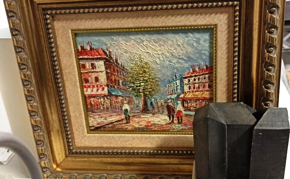
a) Original oil painting to be examined by x-ray radiography.
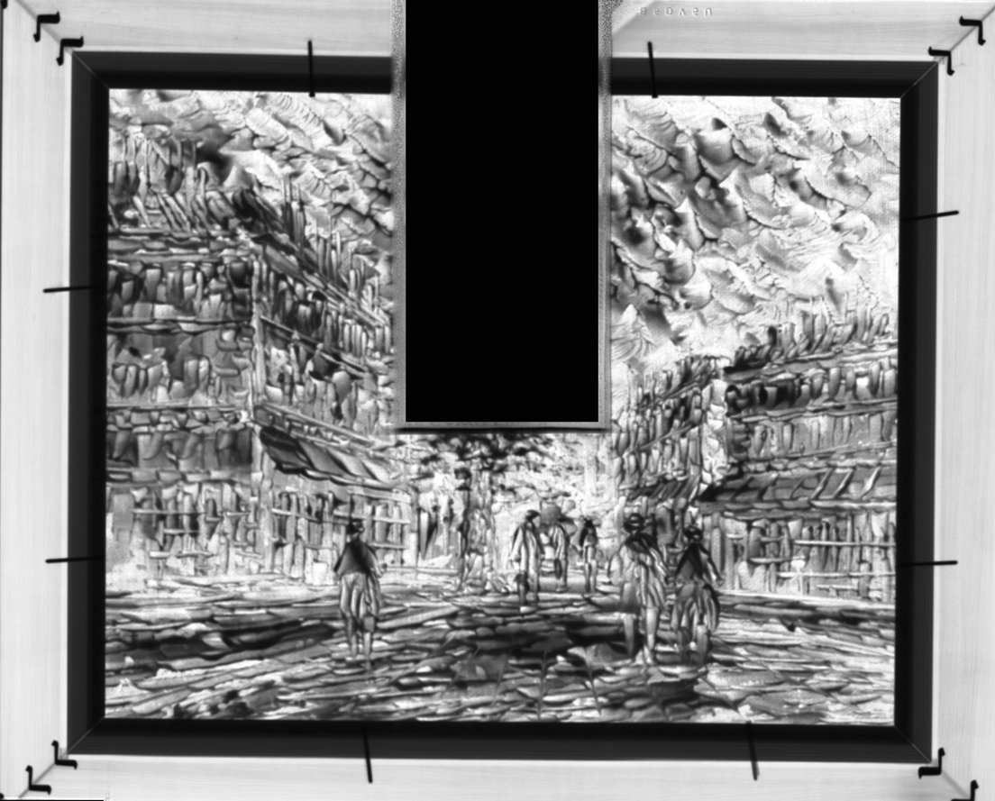
b) High resolution image of the oil panting using a NeutronOptics x-ray camera. Note the absorption of the different pigments, depending on their atomic composition and thickness, and the nails holding the painting in its wooden frame (Prof. Miguel Ángel Respaldiza Galisteo and colleagues, Centro Nacional de Aceleradores, Seville).

Neutron image on a 100 kW Triga reactor X-ray image on a 60kV/3ma/10s x-ray source by Dr Robert Zboray (Penn. State Uni.)
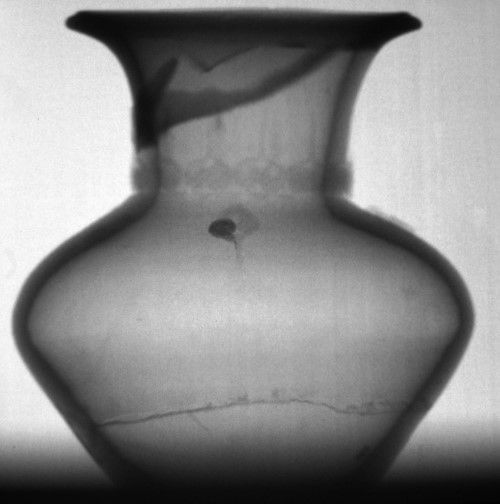
a) High resolution 10s neutron image of an invisible crack in an ancient vase from the 1.3MW Triga reactor at the Thai Institute for Nuclear Technology (Dr. K Sasiphan, TINT)
Touch the mouse on the image to apply imageJ's band-pass filter to emphasise the peaks.
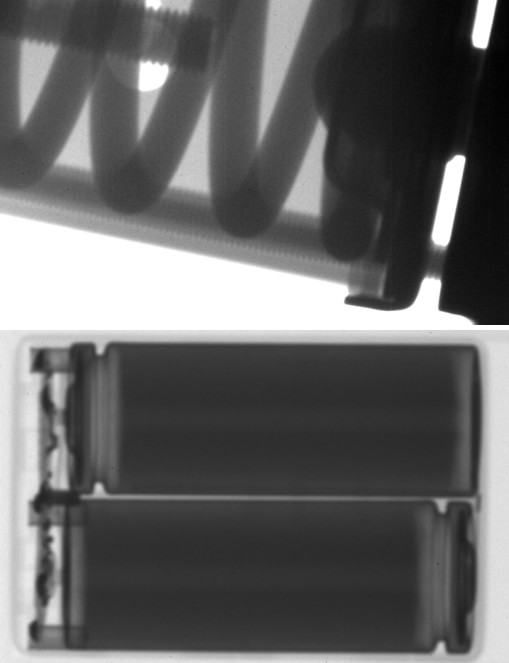
b) High resolution x-ray images of thick objects (a valve and batteries) from only fifty
50-nanosecond pulses from a Golden Eng. 350 kVp pulsed portable x-ray generator
(Prof. Jeffrey King, Colorado School of Mines)
![]()
X-ray backscattering from a twin-CCD Laue camera
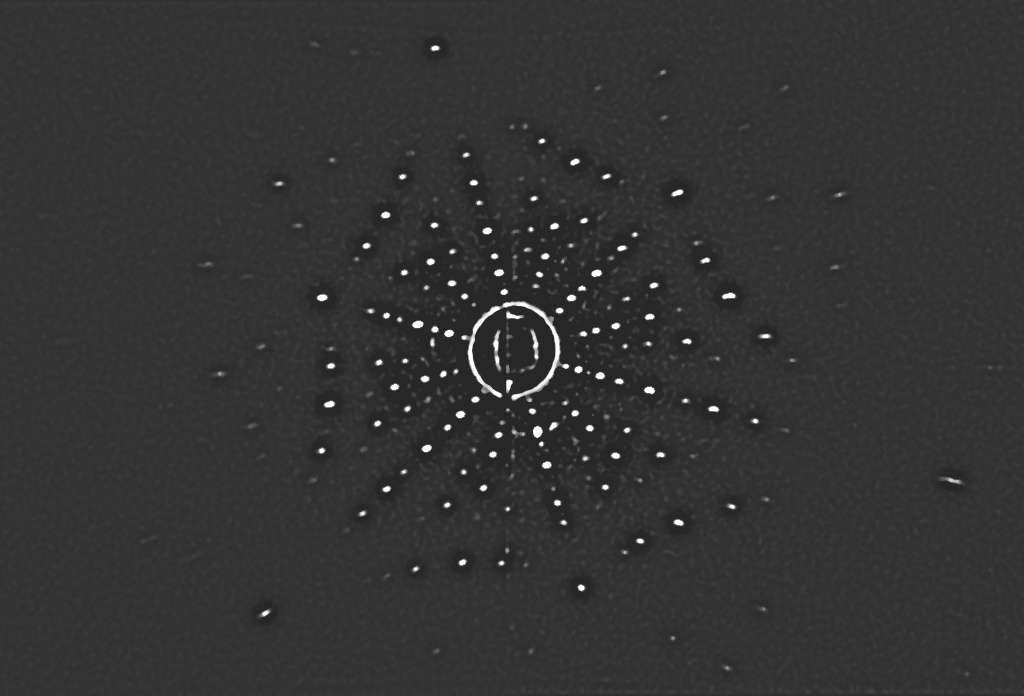
A highly compressed JPEG image of more detailed raw Laue data obtained with our twin high-resolution
Laue camera in 300 secs with 35kV/50mA Cu-anode x-rays
backscattered at 30mm from strongly absorbing Pt(111). Data courtesy of Nakkiran Arulmozhi,
in Prof. Gregory Jerkiewicz's laboratory, the Chemistry Dept., Queen's University, Ontario Canada.
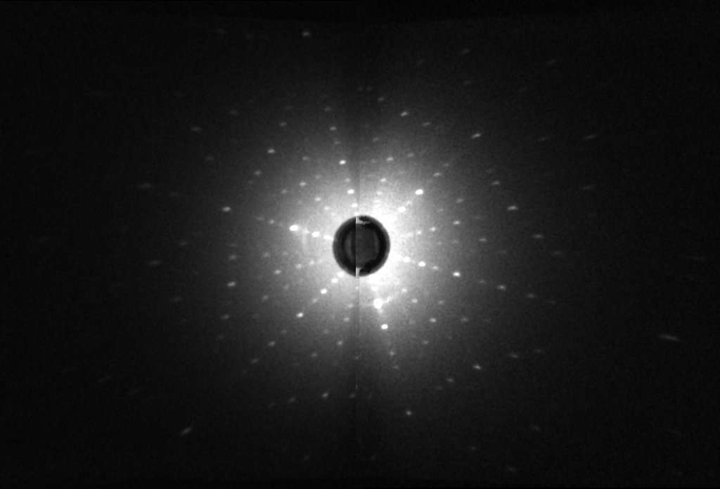
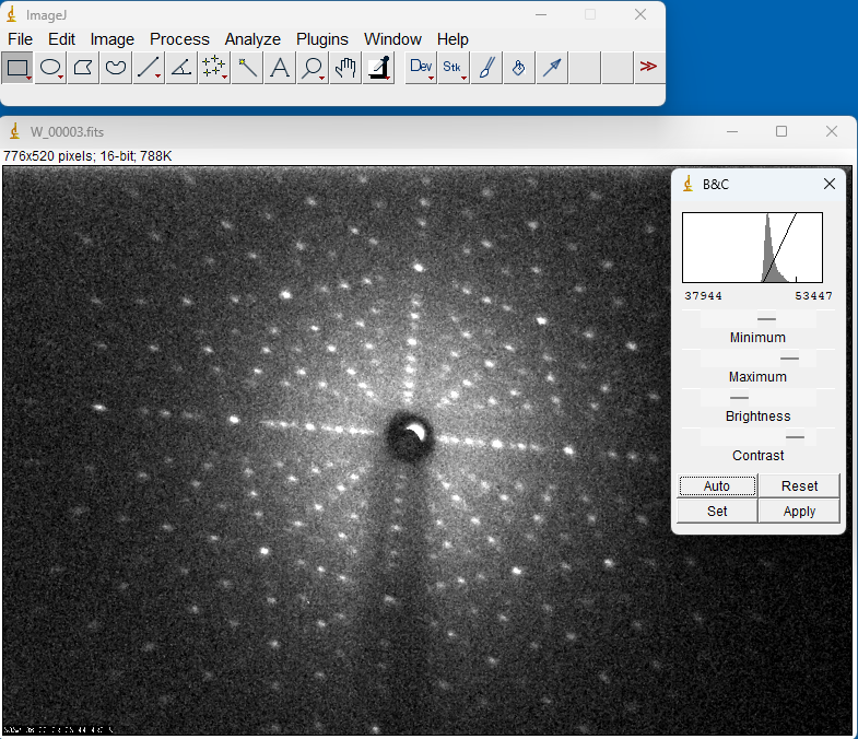
X-ray backscattering with our 1-CMOS Laue camera
Our new 125x100mm 1-CMOS Laue camera is x6 more efficient than our old 1-CCD camera, and a 180x120mm version replaces our old 2-CCD camera. The W-Laue image below was obtained on our 1-CMOS camera in only 30s on a 2kW generator with a W cathode at ~35keV.
Testing a composite neutron monochromator
 A video of the neutron intensity obtained by rocking a composite Pyrolytic Graphite monochromator
through its reflecting angle. The monochromator consists of an array of individually aligned crystals
focusing neutrons onto the sample. The video was recorded on a 100mm camera
two meters behind a "pinhole" aperture at the sample position.
A video of the neutron intensity obtained by rocking a composite Pyrolytic Graphite monochromator
through its reflecting angle. The monochromator consists of an array of individually aligned crystals
focusing neutrons onto the sample. The video was recorded on a 100mm camera
two meters behind a "pinhole" aperture at the sample position.
This shows small mis-alignments of the individual crystals, with different blocks reflecting at
different times as they are rocked through the Bragg angle. These
contributions are superimposed at the sample position, and it is difficult to see the individual mis-alignments.
some composite monochromators reflect only ~50% of the intensity at any one time, and without images like this the problem is not even recognised.
![]()
Dynamic neutron imaging on low flux sources
Above: plastic clock on a 103 n.cm-2.s-1 SANS beam at the Moroccan MINT TRIGA reactor (750kW). 1s exposures recorded on a hand-held video of the CCD monitor (Dr K. Yazid).
Left: boiling water on the Es-Salam reactor in BIRINE (Algeria)
with one of our original small video CCDs (Dr Faycal Kharfi).
A sample in a cryostat within a magnet on D22
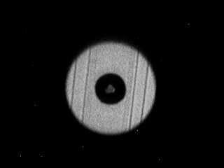
Image with cold neutrons of a sample shielded by an annular cadmium aperture inside a double-walled thin-tail cryostat within a magnet on the small angle scattering machine D22 at ILL Grenoble. The various aluminium walls of the cryostat tail are clearly visible.
First neutrons on SNAP at SNS Oak Ridge
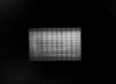
The first neutrons obtained on the SNAP high pressure TOF machine at SNS, Oak Ridge National laboratory, taken from a re-touched photo of the TV screen connected to a NeutronOptics camera. The rectangular structure is an incident beam collimator.
Contrast sufficient to detect a vanadium can
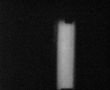
Image of a thin-walled vanadium sample can with cadmium end-caps rotating in a neutron beam on the reactor at Chalk River, Canada. The image is nearly real size.
A weak 2x104.n.cm-2.sec-1 neutron beam
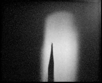
Image of a cadmium pointer on the weak 2x10⁴ n.cm-2.sec-1 neutron beam of IN13 at ILL Grenoble obtained in 20 seconds using the long-exposure option of version 2 of the compact camera. Note the excellent resolution showing a bend in the fine tip of the pointer. This image is larger than the real size of the camera window (60x80mm).
Spatial modulation by Larmor precession (Delft)
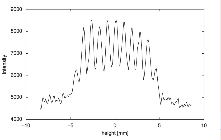
Gaehler has proposed that the intensity of a neutron beam can be spatially modulated using Larmor precession techniques, and this might be used for small angle scattering.
A high resolution neutron detector is needed. The figure shows the horizontally summed intensity, with a modulation wave length of 0.9mm, obtained on SESANS at Delft with a compact NeutronOptics camera. The camera resolution was 0.085mm.
Wim G. Bouwman, Chris P. Duif, Roland Gaehler (2009) Physica B 404 2585–2589.
Incident IRIS beam spreading with wavelength
The incident beam from the IRIS guide tube at ISIS is larger at lower energies (right-hand image) due to the increased angle for total internal reflection at longer wavelengths. Neutron intensity has been mapped to colours using the RoboRealm imaging software.
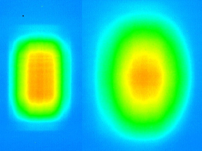
Unfocused and focused monochromator at BARC
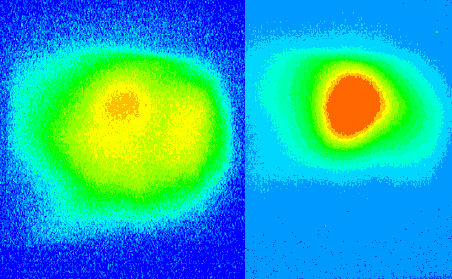
The beam from a flat monochromator (left-hand image) is broader and less intense than that from the focused monochromator at the Bhabha Atomic Research Centre, India. Neutron intensity has been mapped to colours using the RoboRealm imaging software.
Field of view of the compact 80x60mm camera
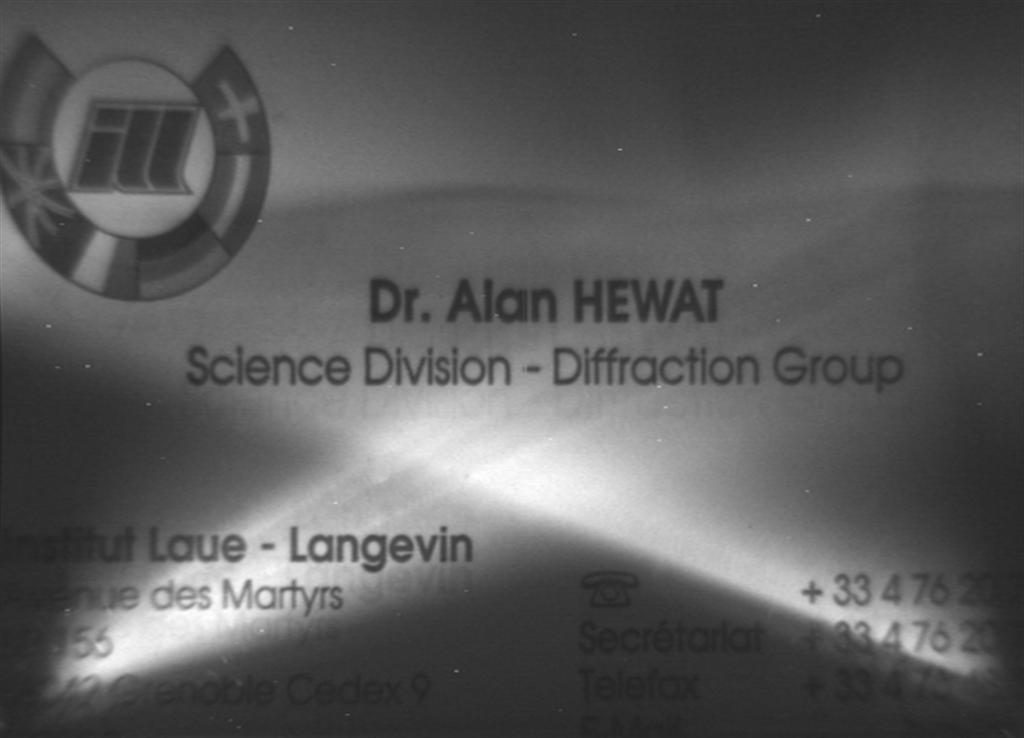
The field of view of the compact camera is about the size of a visiting card.
The photo of a visiting card in place of the scintillator is lit unevenly by a small amount of light leaking in from under the lid, which has been left very slightly open to illuminate the camera for the purposes of illustration. The response of the camera to neutrons is of course uniform, reflecting the uniformity of the scintillator.
The mirror image video has been inverted in real time by the RoboRealm software, and corrections have
also been made in real time for the slight pincushion distortion due to the relatively wide field of view
in the very compact camera body.
![]()